
Imaging diagnosis is one of the pillars of modern veterinary medicine. X-rays, ultrasounds, CT scans, and MRIs provide irreplaceable information about the patient's internal condition. And now, with artificial intelligence, these images are being interpreted more quickly, consistently, and in many cases, more accurately.
How AI analyzes medical images
AI models for imaging diagnosis are based on convolutional neural networks (CNNs) — architectures developed specifically to recognize visual patterns. These models are trained with thousands (sometimes millions) of annotated images from specialists, learning to identify anatomical structures, anomalies, and pathological patterns.
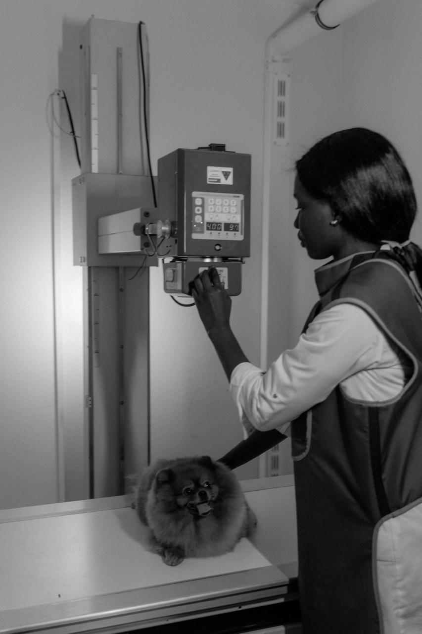
In practice, the veterinarian captures the image with the usual equipment and sends it to the AI platform. In seconds, the system returns a preliminary report highlighting regions of interest and suggesting possible diagnoses.
Applications already available in veterinary medicine
- Musculoskeletal radiology: detection of fractures, dislocations, osteosarcomas, and hip dysplasia
- Thoracic radiology: identification of cardiomegaly, pleural effusion, and abnormal pulmonary patterns
- Echocardiography: automated analysis of cardiac chambers and ventricular function
- Digital cytology: cell classification in digitized slides for neoplasia screening
- Dermatology: identification of skin lesion patterns from photos
Evidence of accuracy
Studies published in journals such as Veterinary Radiology & Ultrasound and the Journal of Veterinary Internal Medicine show that some algorithms achieve sensitivity and specificity above 90% for specific conditions — such as pericardial effusion and hip dysplasia in dogs. In some screening tasks, the models surpass the detection rate of residents and general practitioners.
The veterinarian's role doesn't change — it evolves
AI does not replace the veterinary radiologist. It acts as a second pair of eyes, reducing the chance of lesions going unnoticed due to fatigue or lack of specialization. The final diagnosis remains the responsibility of the professional, who integrates imaging findings with the clinical picture, anamnesis, and laboratory tests.
Real challenges
- Image quality: models trained on high-quality images lose accuracy with inadequate technique
- Data bias: models trained on one population's images may not generalize to other breeds or regions
- Access cost: some platforms still have high costs for small clinics
- Regulation: clinical use of AI in diagnosis still lacks specific regulation in Brazil
What to expect in the coming years
The trend is for AI tools for veterinary imaging to become more accessible, accurate, and integrated into clinic workflows — as is already happening in human medicine with solutions like automated pulmonary nodule detection and diabetic retinopathy screening.
For veterinarians who invest in training and technology today, the return in diagnostic quality and competitive differentiation will be significant.